Short Answer
The dental x-ray bitewing is a quintessential instrument in modern dentistry, playing an instrumental role in maintaining oral health and diagnosing potential complications. The term ‘bitewing’ elicits a sense of curiosity among patients, often prompting one to ponder its precise purpose, safety measures, and the intricate details it reveals about one’s dental anatomy. This article endeavours to elucidate these aspects, presenting an all-encompassing view of bitewing x-rays.
Before delving into the specifics, it’s essential to grasp the fundamental concept of what a bitewing x-ray actually is. A bitewing x-ray is a type of intraoral radiograph that captures the upper and lower teeth in a specific area of the mouth simultaneously. Unlike full-mouth x-rays, bitewings focus on a smaller section, usually encompassing the posterior teeth—molars and premolars—where cavities and dental issues are most frequently identified. This specific positioning aids in illustrating the interproximal surfaces of the teeth, which are often difficult to inspect visually during a routine examination.
Understanding the primary purpose of bitewing x-rays is crucial in appreciating their significance in dental practice. Primarily, bitewings allow dentists to detect caries (tooth decay) that may not be visible during a standard examination. Additionally, they provide information about the bone levels around the teeth, which is imperative for periodontal assessments. The identification of these issues at an early stage can be pivotal in preventing more severe complications that may necessitate extensive treatments, thus acting as a preventive measure in modern dentistry.
Moreover, bitewing x-rays are instrumental during routine dental check-ups, usually conducted annually or biannually. They serve as a benchmark for monitoring dental health over time, enabling practitioners to discern changes that may indicate the progression of decay or periodontal disease. By presenting a visual the practitioner can assess, these x-rays add depth to the diagnostic process.
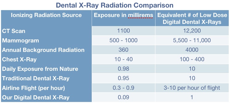
Safety is paramount in any medical or dental procedure, and this is especially true for radiographic imaging. Concerns about radiation exposure are common among patients contemplating dental x-rays. However, it’s crucial to note that bitewing x-rays involve minimal radiation exposure. Contemporary techniques and equipment, such as digital radiography, further reduce this exposure, employing efficient sensors and advanced software to produce images with remarkably low doses of radiation. The amount of radiation emitted from bitewing x-rays is typically comparable to the natural background radiation one might encounter in a single day.
Furthermore, dental professionals adhere strictly to the ALARA principle—an acronym standing for “As Low As Reasonably Achievable.” This principle underscores the importance of minimising radiation exposure while acquiring the necessary diagnostic information. Consequently, it is standard practice for dentists to perform these x-rays only when needed, rather than on a routine basis, ensuring both patient safety and the efficiency of dental examinations.
When a bitewing x-ray is taken, it provides valuable insights and fosters a comprehensive understanding of one’s dental health. The typical interpretation of the x-ray can reveal the presence of cavities, particularly between teeth where they often begin and initially remain unnoticed. Additionally, it can highlight the condition of the bone surrounding the teeth, allowing for the early detection of periodontal disease. This information is indispensable for devising a tailored treatment plan, should any issues arise.
An additional dimension of bitewing x-rays is their utility in monitoring dental restorations, such as fillings and crowns. Regularly taken x-rays offer a visual record, enabling the dentist to evaluate the integrity of these materials and detect any signs of underlying decay or complications that may warrant further attention. This diagnostic approach enhances patient care by ensuring restorations remain functional and effective over time.
While bitewing x-rays serve numerous vital functions, understanding the visual patterns they present can also pique curiosity. A trained eye can discern a multitude of issues from the shadows and contours visible in these radiographs. For instance, radiolucent areas often indicate decay or infection, while dense structures—like enamel and bone—appear radiopaque, showcasing the intricate architecture of the oral environment.
Additionally, bitewing x-rays provide a unique opportunity for patient education. When patients are shown their x-ray images, they can better comprehend their dental health, demographics, and required treatments. This visual evidence fosters open dialogue between the patient and dentist and encourages patients to take a more active role in their dental hygiene practices.
In conclusion, the dental x-ray bitewing serves as an essential component of contemporary dental diagnostics. With its ability to provide detailed visual depictions of dental structures, while ensuring patient safety through minimised radiation exposure, it stands as a cornerstone of effective preventive dentistry. As technology continues to advance, the methods and technologies employed in capturing these radiographs will only enhance their effectiveness, solidifying their indispensable role in fostering optimal oral health. Ultimately, the bitewing x-ray not only assists in diagnosing existing conditions but also acts as a proactive measure in maintaining enduring dental wellness.
FAQ
What is a bitewing x-ray?
A bitewing x-ray is a dental imaging technique that captures images of the upper and lower back teeth to detect cavities and evaluate bone health.
Are bitewing x-rays safe?
Yes, they involve very low radiation exposure and are performed following strict safety guidelines to minimize risk.
How often should bitewing x-rays be taken?
Dentists usually recommend bitewing x-rays once or twice a year during routine dental check-ups, depending on individual risk factors.
What dental issues can bitewing x-rays detect?
They can detect early tooth decay between teeth, assess bone loss related to gum disease, and monitor dental restorations.


Leave a Reply